Concept

Eccentricity — where it appears

How far from the point of gaze a stimulus falls, which decides how many receptors it lands on and which of them are there at all. A few degrees from the fovea the macular pigment has gone, the cones are sparser and the rods are dense, so an observer is a different observer there.

Named by 13 essays across 4 fields — each of them below, with the objects they name alongside it.

Cone density, out from the centre of gaze. Quoted landmarks with logarithmic interpolation between them: 199,000 cones per square millimetre at the fovea, 9,500 at ten degrees — a factor of 21. The shaded bands are the two standard observers' fields. The 2° observer averages over a region whose density falls by 3.3× between its centre and its edge and the 10° observer by 12.4×, which is what "a 10° field" contains and is why the two sets of matching functions are different shapes rather than the same shape scaled.

Colour stops at the edge of sight

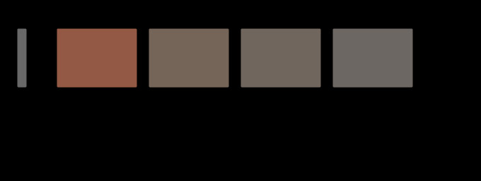
Cone density falls twenty-one-fold between the centre of gaze and ten degrees out, and the three channels give out at three different rates — so a colour difference in the periphery does not merely shrink, it turns. At the exact point of fixation there are no short-wavelength cones at all.
eye · Cones
One colour difference, at four places in the visual field. The same pair of colours — ΔE00 22.0 at the fovea — with each of its three components divided by that channel's own threshold scaling at the stated eccentricity. What is left at 20° is 4.4, and its hue has turned by 26 degrees, because the red–green part is divided by more than the blue–yellow part. The swatches are the predicted colours, drawn where a reader will look straight at them; the figure states a prediction it cannot stage.

A difference has no place

A colour difference formula answers a question about two patches somebody is looking straight at. Move the same pair ten degrees into the periphery and a third of it is left — and it has turned twenty-four degrees of hue, because the three channels give out at three different rates.

difference · Metric
The eye's own drift, and what it does to every spatial frequency. A pattern of f cycles per degree, drifting across the retina at 0.5 degrees a second, arrives at each receptor at f × 0.5 hertz. The curve is the temporal sensitivity at that rate, against the pattern's spatial frequency. Every frequency the eye can resolve stays above a quarter of the temporal peak, and the band of drift speeds for which that holds is 0.02–0.71 degrees a second — with the measured drift inside it. Faster and the finest detail is carried past 60 hertz, where there is no sensitivity at all.

The eye is never still

A perfectly stabilised retinal image disappears within seconds. What keeps the world there is a drift of about half a degree a second between the microsaccades — fast enough to keep the finest detail modulating and slow enough not to carry it past fusion, in a band whose upper edge is at 0.71 degrees a second.

brain · Appearance
A halftone tint away from the centre of gaze, two ways. The upper curve raises the threshold by the E2 rule and leaves the filter alone, which is the multiplication the last phase guessed at. The lower one also moves the cutoff, because eccentricity magnifies the whole spatial scale — implemented as the substitution that makes it exact, a screen of ruling r seen through a filter whose cutoff has been divided by s being a screen of ruling r·s seen at the fovea. The horizontal line is threshold. The screen is visible where you are looking and gone by 3°, while the product prediction has it visible across the whole page.

A tint at the edge of a page

The last phase left this join open and guessed at its answer — how visible a halftone tint is away from the centre of gaze should be the product of two effects it had measured separately. It is not the product. At five degrees the guess is fifty-three times too generous, and by twenty it is out by eight orders of magnitude.

difference · Metric
The drift window, asked about each channel in turn. Every spatial frequency a channel can resolve, drifting at v degrees a second, arrives at f × v hertz; the bar is the range of v over which all of them stay above a quarter of that channel's temporal peak. The luminance band is closed at both ends — 0.018 to 0.71 degrees a second — because its temporal sensitivity has a dip at zero to fall into. The chromatic bands have no slow edge at all, because chromatic temporal sensitivity is low-pass: a stationary chromatic pattern sits at the top of its own sensitivity. The mark is the measured drift, and it is inside all three.

The drift is a luminance mechanism

The eye's own drift was shown to sit inside a band of speeds that keeps every spatial frequency modulating, and the band was quoted as though it were about vision. Asked about colour, it has no slow edge at all — a stationary chromatic pattern needs no eye movement whatever. And a stabilised chromatic pattern is the first thing to fade.

eye · Cones
Where a pooled gain gives out, against where the eye does. The falling curve is how much of a pattern of each spatial frequency a local adaptation pool of 0.5° can see — and therefore how much of it a settled eye can cancel. It is half gone by 0.37 cycles per degree, which is a feature about 2.7° across. The three marks are the acuity limits of the luminance channel and the two chromatic ones. Every one of them is more than an order of magnitude finer than the pool, which is why a stabilised eye loses the fill of a picture and keeps its outline rather than losing the picture.

What a still eye stops seeing

A stabilised image is said to vanish, and nothing in a temporal filter predicts it — sensitivity at zero frequency is a quarter of the peak, not nothing. Give the adaptation gain a size and the answer falls out — fading is a high-pass filter that switches on over a minute, it takes the fill and leaves the outline, and a patch has to be about two degrees across before it goes at all.
eye · Cones
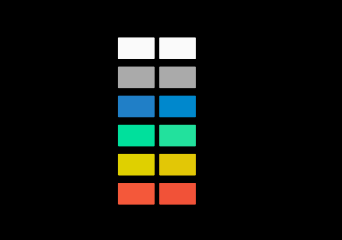
What the same eye reports about one field, in the middle and at the edge. Each row is a uniform field, drawn at the most saturated version of itself this page can show — the percentage is how much of the full stimulus survived, the rest being the adapting light added to bring it inside the gamut. The left patch is what the centre of gaze reports and the right one what 10 degrees out reports, each adapted to the same light as that position sees it. The adapting white comes out identical to 5e-13, because an adapted eye cancels its own filter exactly. Nothing else does, and the largest difference is in the blue. tungsten light is not drawn: it cannot be shown at any useful saturation, and at full strength it differs by ΔE00 1.87.

One person is two observers

The macular pigment is a yellow screen over the fovea and nowhere else, so a cone at the centre of gaze and a cone ten degrees away have different colour-matching functions in the same eye. A match made in the middle comes apart at the edge by six units — and fitting one filter to the gap between the CIE's two standard observers gives a density of 0.40 against a measured 0.35.

eye · Cones
How fine a screen each channel can see. A halftone screen at 35 per cent coverage, 45 degrees, seen by each of the eye's three spatial channels. The bar is the ruling at which it drops below that channel's own threshold. The luminance channel is still seeing it at 55 cycles per degree; the two chromatic ones have lost it by 15 and 10. The dashed line is an ordinary press ruling at reading distance, and only one channel is above it. A halftone is a luminance object, which is why the ink whose dots are nearly the lightness of the paper is the one nobody worries about.

A halftone is a luminance object

The eye's three spatial channels resolve a screen at 55, 15 and 10 cycles per degree, so at any ruling a press actually runs only one of them can see it at all. The corollary corrects a guess already published here — rotating a screen matters more to the chromatic channels, not less — but only at rulings so coarse that nobody prints there.

difference · Metric
The share of itself each change leaves behind, and the smallest is inside the eye. The residual as a fraction of the change rather than as a colour difference, which sorts the census differently. At the top is the macular pigment — the filter in front of the central few degrees of one's own retina — leaving 2.4 per cent of itself. It is a fixed transmittance multiplying the light and the white together, which is as close to a pure gain as anything here gets, and it is why nobody notices they have one.

The filters inside the eye

The macular pigment leaves 2.4 per cent of itself after adaptation — the smallest share of anything in this site's census of light changes, and less than half the next smallest. Fifty years of lens yellowing leaves 7.9 per cent, and the difference between the two says what a gain is actually good at.

eye · Cones
A stabilised grating fades at a rate that depends on which way its bars run. What is left of a 0.3 cycle-per-degree grating, in multiples of its own threshold, as a function of its orientation, at four moments after the image was stabilised on the retina. The first curve is flat to floating point: at the instant the pattern arrives, orientation does not matter, because the filter that carries it has no orientation preference at this frequency. The anisotropy arrives with the fading, reaching ×2.09 after five minutes, and a pool with no axis predicts none of it.

A fading pool has a shape

Giving the local adaptation pool two axes instead of one costs a single parameter and produces a prediction the circular version cannot make — a stabilised grating fades at a rate that depends on which way its bars run. The obvious objection is the oblique effect, and the two act in bands that do not overlap.

eye · Cones
A gap in the wall and a gap in the drive are not the same gap. What the centre of a region settles to under three conditions. With the contour closed it reaches its border's value exactly. Open a 16% hole in the barrier and leave the border signal unbroken and it still reaches it, to 1e-8 — a ring of driven cells encloses the centre whatever the wall outside it is doing, so nothing can escape. Break the signal too and it falls to 0.960. All of what a gap costs is the piece of border that stopped driving, and none of it is the hole.

A gap in the drive, not in the wall

Break the contour around a region and leave its border signal unbroken, and the interior does not move by one part in a million — a ring of driven cells encloses a centre whatever the wall outside it is doing. Break the signal too and the shortfall goes as the square of what is missing. All of what a gap costs is the piece of border that stopped driving.

scene · Scene
The three cone absorptances at two settings of the field size. Solid and dashed are the same construction at the two ends of the CIE's own second observer. The curves are built from one pigment template through its ocular media, which is the same model its population of two hundred eyes is drawn from. The largest difference between the two sets is 16.0 per cent of the peak, and where it sits along the wavelength axis is what decides which stimuli the two observers disagree about — a departure concentrated in the blue is invisible on a sample with no blue in it.

A field size is two changes

The CIE publishes two standard observers and the difference between them is usually described as a field size. What actually differs is a macular pigment the light no longer passes through and a cone outer segment the light no longer travels the length of — two changes, in two places, with different signs and different sample dependence.

eye · Cones
The three cone absorptances at two settings of the macular pigment. Solid and dashed are the same construction at the two ends of two standard deviations of the reported spread. The curves are built from one pigment template through its ocular media, which is the same model its population of two hundred eyes is drawn from. The largest difference between the two sets is 31.5 per cent of the peak, and where it sits along the wavelength axis is what decides which stimuli the two observers disagree about — a departure concentrated in the blue is invisible on a sample with no blue in it.

The macular is a band, not a filter

The lens absorbs everything below 500 nanometres with a long tail; the macular pigment absorbs forty nanometres either side of 460 and nothing else. The two have similar sizes and completely different distributions, and the reason is that one is broad and one is narrow — which is what decides whether a sample is affected at all.

eye · Cones

Named alongside it

The objects these essays reach for when they reach for this one.

ThresholdIndividual variationSpatial frequencyContrast sensitivityOpponent processingAdaptationAssertionStandard observerCone fundamentalsMacular pigmentOrientationTemporal sensitivity

All concepts